Notice
For patients who will undergo surgery
Please carefully read the information provided below regarding the surgical procedure you will undergo:
- You can call our office at 0312 593 41 39 before or if needed.
- Please do not eat or drink anything, including water, from 12 o’clock the evening before the surgery.
- Wash your leg thoroughly with warm soapy water the day before the surgery. Take care not to damage or injure the skin during this process.
- Necessary tests will be performed after you are admitted to the hospital. However, those that have been done beforehand…
Please remember to bring all your laboratory and radiological test results with you.
It is advisable to come to the hospital wearing clothing that you can easily put on over your post-operative bandages.
Please report to the relevant department at the hospital you have been informed about. After this stage, you will be admitted to your assigned room, and the nurses will obtain your initial information.
Please be sure to inform the nurse and our anesthesiologist if you have any allergies or if you have any medications you have used up to that day.
The specialists of the Department of Anesthesia will meet with you before the surgery. During this meeting, you can exchange views on the general anesthesia (narcosis) or spinal or epidural anesthesia (numbing only that area) that will be administered to you. Remember to inform them about the medications you are taking and your allergies during this meeting. You can also receive information about post-operative care. In this meeting, you can get clarifying answers to your questions about how to manage post-surgical pain and other related issues.
Afterwards, Prof. Dr. Mehmet Binnet will meet with you to provide final information and answer your questions.
Your post-operative care
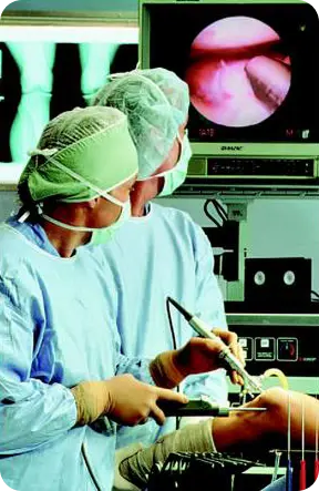
Although surgical treatments are used to solve many problems, you may still experience some limitations in your activities even after recovery. The results of treatments will depend on the degree of injury or damage detected in your knee. If you have undergone an arthroscopy procedure on your knee joint, complete healing will take several weeks, even though the entry points are small and you may feel some pain in the joint where the arthroscopy was performed. To speed up the healing process and protect your future joint function, you will be recommended an exercise and rehabilitation program.
For example, if you injured your knee while running and the joint surface of the weight-bearing part of the knee is damaged, immediate healing should not be expected. Depending on the extent of your problem, you will be recommended a low-impact exercise alternative. Athletes may recover faster than those who only do daily activities. This is because athletes have stronger muscles around the knee than normal individuals, and their recovery can occur more quickly with exercise. However, our main goal is for our patients to return to work, school, or daily activities in a short time. Physical exercise and the rehabilitation process play the most important role in your recovery. A regular exercise program will greatly contribute to your final condition. For athletes or those in good physical condition, returning to sporting activities is possible within a few weeks. However, please remember that the differences in arthroscopy procedures and the varying findings and conditions of diseases mean that the arthroscopy procedure is a personalized process for each patient. The healing process varies according to these characteristics.
However, it is useful to keep the following general characteristics in mind:
- If you feel fullness in your joint after surgery, this is normal. Some of the water injected into your knee during arthroscopy may have remained inside, and this will be absorbed by the body over time.
- An elastic bandage has been applied to your knee and leg to prevent swelling after surgery. You will be informed about how many days after surgery the bandage can be removed.
- It is beneficial to elevate your legs by placing 2-3 pillows under them while you are lying down. As a rule, it is beneficial for your knee to be higher than your heart level.
- You can apply ice to your knee for the first two days. Applying ice for 15 to 20 minutes, five or six times a day, will be beneficial against pain and swelling in your knee.
- Ice should not be applied directly to the skin. The easiest way to do this is to wrap a plastic bag filled with ice cubes in a towel and place it on the area. If you have a bandage, it should be applied over the bandage.
Immediately after the procedure, start moving your foot and toes as soon as possible. This is beneficial for blood circulation. Also, exercises involving contracting and releasing the thigh muscles should be started immediately.
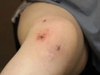
The incisions in the skin are usually closed with stitches; the stitches are removed after ten to fifteen days. During this time, the area where the procedure was performed should not come into contact with water. It is beneficial to clean this area after the surgical site has been wrapped with plastic wrap.
You will be discharged with the medication(s) to be administered, a home program, and the date and time of your follow-up appointment. For necessary information regarding your follow-up, contact Prof. Dr. This will be communicated to you by M. Binnet and his team.
During this period, if your knee swells severely, warms up, or if fluid comes from the wound sites after two days, or if there is discoloration throughout your leg, please contact us as soon as possible. - The exercises that will be given to you after surgery are important for you to return to daily life or sports more quickly. Exercise your knee as much as possible. Tighten and release your thigh muscles for ten minutes every hour, and slightly lift your leg from where you have it straight, hold it in this position for five seconds, then put it back down. This will prevent fluid buildup (knee swelling) in your knee.
- The diligence you show regarding exercises will undeniably shorten the recovery time and allow you to return to work earlier.
- In addition to all the information, if you have any questions, please contact us at 0312 593 41 39
What is arthroscopy?
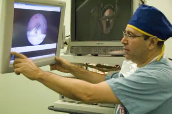
Arthroscopy is a minimally invasive surgical procedure used in the diagnosis and treatment of joint diseases. Arthroscopy allows your doctor to see inside the knee joint in detail through 4.5 mm incisions using a small lens and lighting system attached to a pencil-thin instrument called an arthroscope. The word arthroscopy is derived from Latin, from the roots artros [joint] and skopi [to observe]. Therefore, arthroscopy means “looking inside the joint.” The instrument that allows us to see inside the joint is called an arthroscope. Arthroscopy uses a lens and video camera system connected to a fiber optic light source. To observe the inside of the joint, the optical system of the arthroscope is inserted into the joint through 0.5 cm incisions. Arthroscopy is essentially an optical system. A camera attached to the end of the optical system, called a scope, allows the entire inside of the joint to be viewed on a monitor. Because the images inside the joint are magnified 6-10 times, a very detailed examination of all the structures inside the joint is possible. Other surgical instruments can be sent through another incision to repair damaged tissues in the knee, depending on the arthroscopy images and diagnosis. Arthroscopy can be performed in a shorter time compared to other surgeries. Images can be recorded and photographs can be taken. In this context, the inside of the joints is observed through minimal and very small incisions, without making the large incisions required in open surgical treatment methods.
Furthermore, arthroscopy allows for a more complete examination because it can visualize areas inaccessible during open surgery.
Knee arthroscopy was first performed in 1960. Until the 1980s, arthroscopy was used solely for diagnostic purposes, but with advancements in technology and experience, it has become an undisputed treatment method today. Improvements in arthroscopy instruments, along with the development of higher-resolution cameras, have made the procedure more effective in diagnosing and treating knee problems. Today, arthroscopy has become the most frequently performed orthopedic surgical procedure. For example, in the United States, more than 1.5 million arthroscopic procedures are performed annually.
For large joints such as the knee and shoulder, 4 mm diameter arthroscopes are used, while for smaller joints, 1.9-2.7 mm diameter ones are used. By entering through these small incisions, diseases of the articular cartilage, menisci, ligaments, the membrane lining the joint, and fractures and dislocations affecting the joint can be treated. Today, arthroscopy is used more as a treatment tool (arthroscopic surgery) than a diagnostic tool. For this, in addition to the imaging system, specially developed mechanical or motorized instruments with diameters of 2.7-6.5 mm are used. Depending on the definitive diagnosis made during arthroscopy, arthroscopic surgery is performed in the same session.
The main benefit of the technique is seen after the surgery. Because the joint is not opened, physical therapy and rehabilitation can begin earlier and are easier. Since arthroscopic surgery is performed through very small incisions, it is the method that causes the least damage to normal tissues. Patients experience much less postoperative pain compared to open surgical procedures. Thus, the patient recovers faster and returns to an active life sooner. The ability for athletes to return to sports sooner is a great advantage.
If sufficient exercise is performed after arthroscopy, the risk of developing limited range of motion in the joint is negligible compared to open procedures. Similarly, problems such as infection and thrombophlebitis are less common. For all these reasons, the recovery period after arthroscopic surgery is shorter and more comfortable.
HOW IS ARTHROSCOPY PERFORMED?
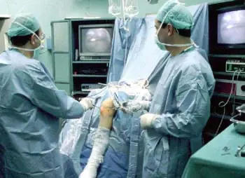
Arthroscopic surgery requires operating room conditions and anesthesia. Diagnostic arthroscopy can be performed with local anesthesia. Surgical arthroscopy often requires general or spinal anesthesia. A 0.5 cm incision is made to view the inside of the joint. Several more incisions may be necessary for diagnosis and treatment. The instruments required for the surgical procedure are inserted into the joint through a second incision. The surgical procedure is performed while viewing the inside of the joint on a monitor. The entire arthroscopy can be video recorded if desired. After the procedure is completed, a drain may be placed to remove accumulated fluid from the joint. This drain is often removed during the next dressing change. Mild pain relievers are usually sufficient after arthroscopic surgery. Depending on the type of procedure performed, the hospital stay varies between one and two days; generally, it is one day, except for patients undergoing cruciate ligament or patellar dislocation repair.
No cast is applied after arthroscopic surgery; in some cases, knee braces allowing for controlled movement are used. After some procedures, crutches may be used for 3-4 weeks to avoid full weight-bearing on the operated leg. You will be informed about which movements and exercises are permitted after arthroscopy and how to care for the wound. Assistance will be provided regarding suture removal and the rehabilitation program during follow-up appointments. The recovery time may vary depending on the extent of the procedure.
Complications after arthroscopy are uncommon. However, although arthroscopy is a minimally invasive procedure, there is a slight possibility of postoperative swelling or blood accumulation.
The development of infection or inflammation is much less frequent compared to other procedures. Arthroscopy is performed with protective measures in place for these issues. You can obtain more detailed information on these matters during your consultation with Prof. Dr. M. Binnet and ask any questions you would like clarified.
WHEN IS ARTHROSCOPY NECESSARY?
The diagnosis of joint diseases is made with the help of a good history, physical examination, direct radiographs, and laboratory tests. Computed tomography and magnetic resonance imaging may be used when necessary. Despite all this, arthroscopic examination may be performed in problems where diagnosis is difficult. However, nowadays, arthroscopy is widely used in the problems listed below. Because arthroscopy is frequently used in the treatment of patients with joint problems. The procedure is performed on the knee, shoulder, ankle, wrist, elbow, and hip joints, respectively.
IN THE KNEE JOINT:
The most common problems in the knee are:
Meniscus tears,
Ligament tears, especially anterior cruciate ligament,
Articular cartilage damage,
Free bodies (joint mice),
Inflammation and enlargement of the knee joint capsule (synovitis); usually due to rheumatic conditions. Arthrosis or Osteoarthritis
The main procedures that can be performed arthroscopically (minimally invasively) in the knee include:
Removal of torn meniscus fragments,
Suturing of some meniscus tears,
Anterior and posterior cruciate ligament repairs,
Early treatment of osteoarthritis,
Osteochondritis (cartilage detachment or joint mice),
Fractures involving the knee joint,
Cartilage transplants,
Correction of patellar (kneecap) axis,
Treatment of patellar dislocations,
Drainage of joint inflammations,
Removal of diseased joint membrane (synovectomy),
Restoration of movement restrictions caused by accidents or illnesses,
Removal of benign intra-articular tumors and cysts can be performed arthroscopically or with arthroscopy assistance.
SHOULDER:
Treatment of muscle impingement and recurrent shoulder dislocations,
Interventions for cartilage and muscle tendon diseases within the shoulder joint,
Early treatment of osteoarthritis,
Synevectomy in rheumatic diseases [removal of the thickened membrane lining the joint],
Restoration of shoulder range of motion restrictions,
Removal of intra-articular free bodies can be performed arthroscopically or with arthroscopy assistance.
ANKLE:
Intra-articular fractures, osteochondritis [cartilage fragmentation and joint mice],
Meniscoid lesions [tissue impingements occurring after recurrent sprains],
Early osteoarthritis,
Arthroscopic diagnosis and treatment of rheumatic diseases can be performed.
WRIST:
Treatment of intra-articular fractures,
Relief of nerve compressions,
Treatment of ligament tears between the wrist bones,
Treatment of articular cartilage damage,
Correction of TFCC [special cartilage cushion within the joint] tears can be performed arthroscopically.
ELBOW:
Treatment of osteochondritis [cartilage detachments and joint mice],
Removal of loose bodies,
Filing down bone spurs that restrict movement,
Synevectomy [rupture of the thickened membrane lining the joint] in rheumatic diseases,
Removal of benign intra-articular tumors and cysts can be performed arthroscopically or with arthroscopy assistance.
Ankle sprains and first aid measures
Ankle ligament sprains are the most common injuries in sports and daily life. International studies report that 27,000 ankle ligament injuries occur every day. In other words, one in every 10,000 people sprains their ankle daily. At least half of these are serious sprains. It is essential to take these injuries seriously and take the necessary precautions.
These injuries are also common in athletes. 45% of basketball injuries, 30% of football injuries, and 25% of track and field injuries occur in the ankle joint. 75% of ankle injuries are ligament injuries, and 85% of these are caused by inward twisting of the foot, resulting in injury to the lateral ligaments. If protective measures are not taken after an injury in athletes, sprains frequently recur. This results in deterioration of the cartilage in the joint. The consequence is a long-term interruption of active sporting life.
Anatomically, the ankle is the joint formed by the talus (ankle bone), formed by the two long bones of the leg, the tibia (thick) and fibula (thin). There are three ligaments on the lateral side and a single, strong deltoid ligament on the medial side. As previously mentioned, the weaker lateral ligaments are much more frequently injured. Medial collateral ligament injuries are less common but more serious and take longer to heal.
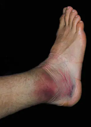
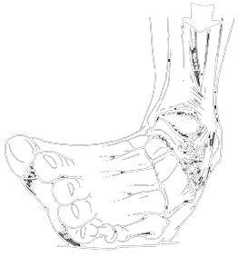
A sprain refers to damage to a ligament. In a mild injury (grade 1), there is no tear in the ligament, only stretching or elongation of the fibers. In a grade 2 (moderate) injury, some fibers of the ligament are torn. In a grade 3 (severe) injury, there is a complete rupture of the ligament. The most frequently injured ligament in the ankle is the lateral anterior ligament (TIA). A severe, or grade 3, sprain indicates rupture of both the lateral and medial ligaments, resulting in joint laxity.
In ankle sprains, the first step is to determine the severity of the injury. It may be harmful to treat a mild sprain with cold application and bandaging, allowing the athlete to continue playing. If examination reveals significant joint laxity and the athlete experiences severe pain and limping when attempting to run, it means they will be unable to continue their sporting activity or current activity. If the ankle swells immediately and restricted movement develops, the likelihood of intra-articular bleeding is high.
Most sprains primarily involve immediate treatment with rest, cold application, compression with an elastic bandage, and elevation of the leg. Crutches and light splints are beneficial to protect the injured area from further damage. Cold or ice application should be done immediately to reduce swelling and promote faster healing. This is done by wrapping an ice pack in a towel and applying it for 15 or 20 minutes every hour. This may take 1-3 days depending on the situation. A definitive diagnosis should be made during this time. Weight-bearing should be avoided on the affected leg as long as pain persists. Rehabilitation can take 1-4 weeks depending on the severity of the injury.
After returning to sports, the risk of re-spraining is high for a period of time. Generally, a second sprain is worse than the first, and a third is worse than the second. Therefore, using a bandage or ankle brace for 6 weeks after the injury may be appropriate to ensure adequate healing. Ankle sprains are not serious injuries, but they require rehabilitation for effective treatment. This is not an injury that can be treated with just rest, bandaging, and medication. If treated that way, chronic ankle laxity can often develop.
Back pain
Back pain is one of the leading health problems causing discomfort and preventing people from working today. In this context, 65% of people experience back pain at some point in their lives. Furthermore, one-third of rheumatic complaints originate from the lower back. In addition, diseases of neighboring organs, other diseases affecting the entire body, and congenital anomalies can all cause back pain.
BACK PROBLEMS RESULTING FROM STRAIN OR INJURY
Back problems resulting from traffic accidents, falls, lifting heavy loads, sudden and awkward movements of the lower back, and excessive strain on the lower back during sports activities without training are included in this group. Both direct and indirect injury mechanisms, depending on the severity of the injury, can lead to conditions ranging from sprains to ruptures, and even spinal fractures and dislocations.
Among these, the largest patient group consists of problems related to lumbago, sciatica, and herniated discs. Most are caused by lifting heavy objects or sudden awkward movements. Women’s daily lives, which involve constant movement and sometimes heavy lifting, increase their risk of developing these types of back problems. The pain is described as a sudden, sharp sensation in the back, like something snapping, when lifting something heavy. The person freezes in place and says their back is stiff. Muscle spasms are often present. Movements are painful. Coughing, sneezing, and straining can worsen the pain. As the symptoms progress, pain radiating to the leg, tingling, numbness, and loss of reflexes may occur.
In sciatica, there is localized pain along a unilateral nerve in the leg. Treatment for all these problems involves rest, medical treatment (medication), and physical therapy agents.
However, from a public health perspective, preventive measures are more important than curative services. Naturally, this principle applies to women’s risk factors in their home and professional activities. The basis of preventive measures is to avoid incorrect movements that may occur when sitting or lifting something from the ground.
The main causes of back pain are addressed within various disease groups. Determining whether there are back problems developing after congenital anomalies is the first step in this regard. Congenital anomalies causing back pain generally originate from the anatomical structure of the spine and are associated with problems in the bone structure. Although the condition is congenital, complaints and findings usually arise in middle and old age due to strains, progressive arthritic changes with age, or injury.
Pain is generally the most common symptom. In addition, varying degrees of limitation in some movements of the spine, stiffness and tenderness in the back muscles, and changes in the normal curvature of the spine (decrease or increase) constitute the main clinical findings. Diagnosis and treatment, especially in diseases with curvature of the spine, are determined through examinations performed under the supervision of a physician.
MECHANICAL CAUSES OF LOWER BACK PAIN
This group, which constitutes the most important part of lower back pain, consists of lower back pain that does not have the severe symptoms specific to the problems presented above, but is caused by external stresses on the surrounding soft tissues.
Abnormal loading on the soft tissues of the lower back: Obesity, pregnancy, the habit of constantly wearing high heels, leg length discrepancies, hip and knee problems, and changes in the body’s weight distribution such as flat feet place more load on the lower back than normal, creating a زمینه for the development of a number of lower back complaints. Naturally, in treatment, it is necessary to minimize the causes that cause the lower back problems before addressing the problems themselves.
Another factor that can be held responsible in this regard is weakness and insufficiency in the abdominal and lower back muscles. A weak muscle structure reduces the body’s natural defenses and cannot prevent lower back problems. Therefore, it is necessary for women, especially those in active working lives, to incorporate exercises that strengthen the lower back and abdominal muscles into their daily lives. A regular morning exercise habit is a preventive measure against lower back problems. Initially, the intensity of exercises is light, and this intensity is gradually increased over time. The aim of the exercises is to reduce lordosis, correct posture, and maintain mobility. This also helps prevent poor posture habits.
It should be remembered that in other diseases causing back pain (inflammatory, rheumatic, metabolic, tumors, circulatory-related, or others), treatment is directed at the underlying disease.
PREVENTIVE MEASURES FOR BACK PROBLEMS
- One should sleep on a flat mattress. The mattress should not be unnecessarily hard. Foam or spring mattresses should be such that the person can easily adjust to a proper position upon getting up. When lying on their side, the legs should be bent at the hips and knees.
- Soft furniture and deep sofas are not recommended. Sit upright in a firm chair with hips and knees at a 90-degree angle and feet flat on the floor.
- Car seats should be firm. Sit close to the steering wheel to avoid straining your legs.
- Do not lean too far forward or backward while sitting. Sudden turns to reach for something are dangerous.
- Keep your back straight while standing. To prevent increased lordosis in the lower back, place a footrest under one foot (if standing for a long time).
- Women should not wear heels too high.
- Do not lift excessively heavy objects. Keep objects close to your body while lifting. When getting up from the ground, squat down for support and push from waist level.
- Exercises should be stopped before reaching the fatigue limit.
However, regular exercise should be done every day.
- Stairs should be taken up and down one step at a time.
- Those with back pain should not sit for long periods. Also, chairs with back support should be preferred.
How does the knee joint work and why is it frequently injured?
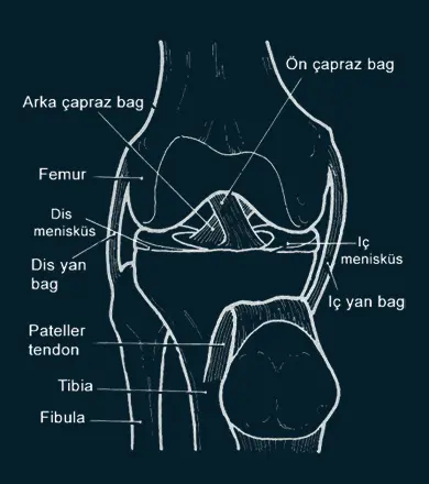
The knee is the largest joint in our body, and it’s important to remember that due to its anatomical features, it’s also the most easily injured. The joint consists of the lower end of the femur (lower leg bone), the upper end of the tibia (shin bone), and the patella (kneecap) which extends along the groove at the end of the femur. These two bones, structurally different, maintain the continuity and stability of the joint thanks to the anterior and posterior cruciate ligaments and the medial and lateral collateral ligaments. The stability of the femur-tibia joint is maintained by the integrity of these anatomical structures. Strong calf muscles provide strength and control to the knee. If the muscles surrounding the knee are not strong enough, the likelihood of knee joint injury increases.
The surfaces where the femur, tibia, and patella meet are covered with articular cartilage, which acts as a cushion between the bones and allows for smooth movement. Semicircular rings of cartilage tissue, called the lateral and medial menisci, act as shock absorbers and provide stability against loads or impacts.
The knee bones are covered by a thin, smooth joint capsule. A thin synovial membrane surrounding this capsule secretes a special fluid that lubricates the knee and reduces friction to almost zero in a healthy knee. This thin, fatty tissue is the source of the joint fluid.
Cartilage injuries
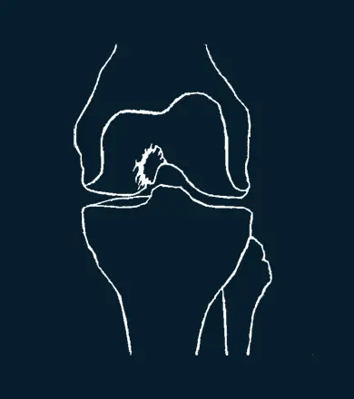
The shiny white structures that form the surfaces of the bones facing each other in a joint are cartilages. Their most important function is to bear weight, and they also provide lubrication to the joint.
Knee joint cartilage is an average of 2 to 4 mm thick, non-vascular, and nerve-free structure. Therefore, its healing capacity is very limited. It is very difficult for a problem to resolve itself spontaneously.
Cartilage injuries can occur directly or as a result of other joint problems, such as in traffic accidents, falls on the knee, or impacts. Symptoms can be mild or severe depending on the severity of the cartilage injury. Common symptoms are swelling and pain in the joint. Symptoms worsen with daily activities or sports. Pain also tends to increase with activities that put more strain on the knee, such as climbing stairs. If the complaints persist, diagnosis and treatment are necessary. Examination and radiological tests provide conclusive information. In recent years, cartilage problems have been more easily and clearly identified thanks to MRI and arthroscopy.
The most valuable diagnostic method is arthroscopy. The ability to visualize the problem in the cartilage, determine its localization, boundaries, dimensions, and depth, and most importantly, to begin treatment in the same session, makes arthroscopy indispensable.
Regardless of the treatment method chosen for cartilage lesions, the location, depth, and boundaries of the problem must first be very well determined, staged according to classification, and then treatment should be initiated according to the chosen method. Based on this data, non-surgical and surgical treatments can be considered. Today, surgical treatments, depending on the size and characteristics of the problem, include cartilage surface shaving, abrasion arthroplasty, subchondral drilling, microfracture, osteochondral autografting, osteochondral allografts, chondrocyte culture, and transplantation. Advances in these methods increase the percentage of hyaline cartilage in the repair tissue.
The primary goal in the treatment of cartilage lesions is undoubtedly for the newly formed cartilage surface to consist entirely or almost entirely of hyaline cartilage. For better post-surgical recovery, early mobilization and avoiding weight-bearing for 4-8 weeks, depending on the procedure performed, should be followed.
Meniscus tears
The menisci are crescent-shaped fibrocartilaginous structures (composed of connective tissue and cartilage) that cover 1/2 to 1/3 of the articular surface of the upper end of the tibia (shinbone) in the knee joint. They consist of collagen fibrils arranged in a way that provides elasticity to withstand compressive forces. The peripheral edges of the menisci are convex and fixed to the inner surface of the joint capsule. The medial meniscus is also attached to the medial collateral ligament of the knee joint. The medial edges of the menisci are concave, thin, and free. Their lower edges are straight, while their upper edges are concave. The part attached to the joint capsule is vascularized, while the other parts are avascular. The medial meniscus is C-shaped, and the lateral meniscus is C(0) shaped, covering 2/3 of the tibial flat surface. The lateral meniscus is more mobile than the medial meniscus. Due to these anatomical structural features, the medial meniscus is more prone to injury than the lateral meniscus.
Functions of the Menisci:
For the normal function of the menisci, the normal function of the knee joint is required.
- Ensuring joint stability,
- Shock absorption function (absorbs forces applied to the knee joint, acts like a shock absorber),
- Ensuring surface compatibility of the joint,
- Ensuring even distribution of joint fluid (lubrication),
- Preventing compression of the synovial membrane (joint membrane),
- Breaking down excessive extension and flexion of the knee joint,
- Ensuring the transmission and passage of loads applied to the joint,
- Reducing contact stress by increasing the joint surface area.
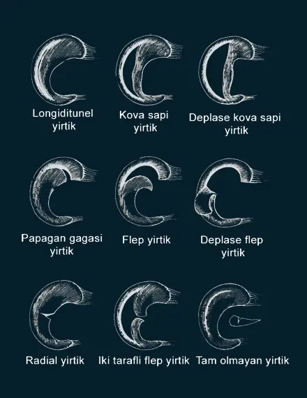
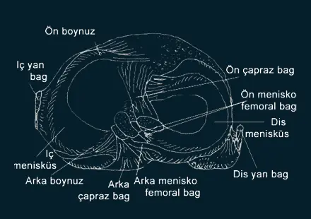
Meniscus Movement:
While following the tibial planes during knee flexion and extension, the menisci follow the femoral condyles during rotation. Menisci are usually injured during rotation of the knee joint. Due to their elasticity and attachment points, the menisci do not move towards the center of the joint.
CLINICAL EXAMINATION
The examination should cover the entire lower extremity (leg). A good and careful history taking, physical examination, and standard radiographs, along with NMR and arthroscopy, are used to make the diagnosis.
Symptoms of meniscus tears are grouped into two categories:
- Group with locking and clear diagnosis,
- Group without locking and difficult to diagnose.
Locking usually occurs in longitudinal (longitudinal), bucket handle tears of the medial meniscus. Locking is when the knee remains in varying degrees of flexion and does not move at all.
Findings to watch out for in the group where diagnosis is difficult without locking:
- Feeling of emptiness
- Swelling (effusion) in the knee joint
- Weakness (atrophy) in the muscles of the anterior thigh and knee, tenderness on pressure in the inner and surrounding area of the knee joint. If a definitive diagnosis cannot be made with these findings, then DIAGNOSTIC TESTS that favor meniscus tears are applied. The results of these tests can give us clues about a meniscus tear. To reach a definitive diagnosis, diagnostic errors can be reduced to below 5% with radiological techniques such as standard X-ray images, computed tomography (CT), and magnetic resonance imaging (MRI). Arthroscopic intervention offers 100% definitive diagnosis and treatment possibilities.
TREATMENT
Meniscus tears are treated conservatively (non-surgically) and surgically (with surgery). In conservative treatment, if the tear is acute (3 weeks), located in the vascularized area of the meniscus, stable with undisplaced edges, and the length of the peripheral tear is less than 15 mm, conservative (non-surgical) healing is possible.
For tears outside these conditions, arthroscopic intervention provides definitive treatment. In peripheral tears (in the vascularized area), the meniscus can be repaired by suturing it back to the torn area. In tears in the non-vascularized area, partial or subtotal meniscectomy is performed, meaning the torn portion is removed. In recent years, with the increasing prevalence of organ donations, meniscus transplantation from a cadaver to a knee in need is also one of the treatment options.
After arthroscopic intervention for meniscus tears, rehabilitation begins immediately. This rehabilitation includes initiating isometric isotonic exercises for the quadriceps (anterior thigh muscles) and hamstrings (posterior thigh muscles). If peripheral (circumferential) meniscus tears are repaired with sutures, you can return to sports after 6 months. In cases of partial meniscectomy, you can return to sports within 2 weeks after good rehabilitation.
Anterior cruciate ligament rupture
ANTERIOR CRUCIATE LIGAMENT INJURIES
The anterior cruciate ligament (ACL) is one of two ligaments located in the middle of the knee joint that cross each other. A rupture of this ligament is the most common serious ligament injury in the knee. Due to the rapidly increasing number of people of all ages participating in sports, the frequency of ACL injuries is also increasing. According to another study from the USA, 250,000 patients are diagnosed with ACL tears every year. This study included both acute and previous tears. If there is a previously existing tear, it is referred to as “ACL insufficiency.” Considering only sports like football and skiing, the frequency of injury is even higher. 70% of acute ACL tears occur during both professional and recreational sports activities.
SYMPTOMS OF ACL RUPTURE
Someone with an ACL rupture will usually experience knee swelling within a few hours following a rotational injury to the knee. The swelling is caused by bleeding into the knee joint from the torn ligament. A clicking sound may also be heard from the knee. Approximately 70 out of 100 athletes with knee swelling after injury should be suspected of having an ACL tear. It is important to determine the nature of the injury by questioning the patient. Most injuries occur when the knee and body are forcefully rotated to one side while the foot is firmly planted on the ground, and often there is no impact involved. An ACL tear can also occur from a kick to the inside of the leg, forcing the knee outwards. It is also important for the patient to say that their knee “twisted” and that they heard a “clicking sound” at the time of the trauma. An athlete with an ACL tear cannot continue the game despite their best efforts and potential strain.
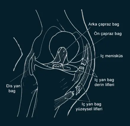
DIAGNOSIS
There are other structures, both inside and outside the knee joint, that may be injured. Therefore, a comprehensive examination of the knee should be performed first. Then, special tests are performed to understand the integrity of the ACL. These tests can reveal an ACL tear. Diagnosis is easier in older tears or ruptures. After the examination, X-rays of the knee should definitely be taken. Fractures or cracks in the bones forming the joint will be visible on the X-ray. A bone fragment that has broken off from the joint surface and fallen into the joint may be seen. Also, the ACL can sometimes detach from its attachment point along with a bone fragment. MRI is useful in showing other possible injuries in ACL ruptures. Meniscus tears are common with ACL tears (40%-60%).
TREATMENT
Several factors must be considered to decide whether surgery is necessary; age, degree of laxity in the knee, the patient’s activity level, and concomitant injuries are the most important criteria for this decision. The primary goal of surgery is to prevent recurrent knee dislocations that may occur later. These frequent knee sprains further increase knee laxity, leading to meniscal tears, cartilage damage, and ultimately, early osteoarthritis. While age is an important factor in deciding on surgical treatment, the individual’s physiological age and activity level are even more crucial. Middle-aged and older individuals are increasingly engaging in sports. Regardless of age, if a person persists with a high activity level, they are a candidate for surgery. Surgical treatment is also considered if knee dislocations and instability occur during daily activities.
Most patients with ACL tears can participate in low-risk activities such as cycling, swimming, and rowing. However, individuals with ACL tears should be warned against high-risk sports. Football, basketball, and volleyball are high-risk sports. The more jumping and sudden turns involved in sporting activity, the more dangerous it is for a knee without an ACL.